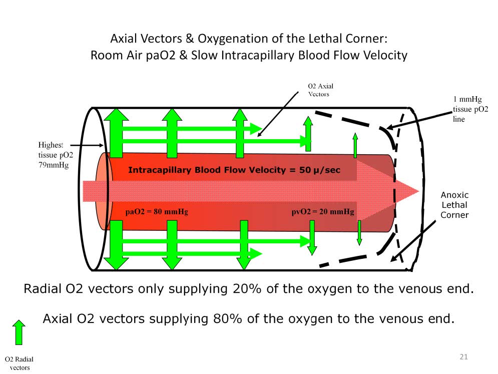
During low flow states axial oxygen vectors may supply up to 80% of the oxygen entering the venous-end tissues of the Krogh cylinder.
When intracapillary blood flow velocity decreases (the cardiac index decreases), the hemoglobin releases more oxygen than normal resulting in a decreased venous hemoglobin oxygen saturation. The radial gradient of oxygen in the venous-end of the cylinder decreases and a lethal corner forms. At the same time, due to the decrease in the venous-end tissue oxygen concentration, the axial gradient increases and more oxygen moves from the arterial-end tissues to the venous-end tissues in an attempt to compensate for the reduced venous radial gradient. In this reduced blood flow scenario the source of oxygen entering the venous-end tissues is only 20% from radial vectors and 80% from axial vectors. That’s not to say that adequate amounts of oxygen are entering the venous-end tissues during low blood flow. Rather that whatever limited supply of oxygen is coming into the venous end comes mostly from axial vectors.
Perfusion Theory is an educational platform for the Oxygen Pressure Field Theory (OPFT). August Krogh’s theoretical concept of the oxygen pressure field is explained and then applied to clinical applications in perfusion practice.